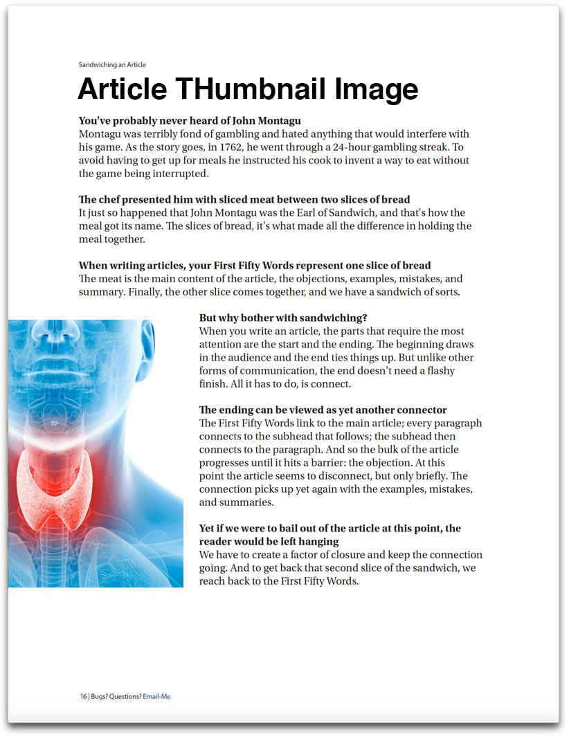
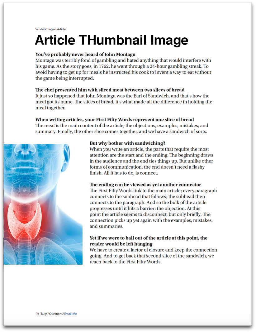
THYROID blood test BASICS
What you can expect when you begin your Defy Medical program.
The first signs of thyroid disorder are typically identified from symptomology and blood screening of Thyroid Stimulating Hormone (TSH) with other thyroid hormone biomarkers, including free thyroxine (T4, free).
Thyroid Stimulating Hormone (TSH), Serum
- TSH is used as a first line screening tool to assess thyroid disease. Additionally, health care providers check TSH to monitor disease progression and treatment
- TSH is high in primary hypothyroidism
- Low TSH occurs in hyperthyroidism
- Evaluation of therapy in hypothyroid patients receiving various thyroid hormone preparations although free T3 should also be evaluated along with patient’s symptoms
- Range: 0.450−4.500 μIU/mL (>10 yr old)
- Methodology: Electrochemiluminescence immunoassay (ECLIA)
T4 (Thyroxine), Total, Serum
- Used for the diagnosis of hypothyroidism and hyperthyroidism
- Free T4 is usually preferred instead of measuring total
- Reference Range: 4.5−12.0 μg/dL
- Most physicians order serum free T4 instead of serum total T4
- Free T4 will provide a better evaluation of bioavailable thyroxine since it tests hormone that is not bound by proteins
T4 (Thyroxine), Free, Serum
- Measurement of circulating thyroxine not bound to proteins (TBP)
- Reference Interval: 0.82−1.77 ng/dl (>19 yr old)
- Methodology: Electrochemiluminescence immunoassay (ECLIA)
- The thyroid gland produces and secretes T4, otherwise known as thyroxine. Proteins bind to T4 and carry it throughout the bloodstream.
- Once in the tissues, T4 is released from the proteins and is now free to convert into the more active form called T3
- Many physicians believe that measuring free T4 is a more sensitive test for thyroid hormone production
Reverse T3 (Triiodothyronine), Serum
- Labcorp Reference Range: 9.2-24 ng/dL
- Methodology: Liquid chromatography/tandem mass spectrometry (LC/MS-MS)
- The rT3 level tends to follow the T4 level: low in hypothyroidism and high in hyperthyroidism
- Increased levels of rT3 have been observed in starvation, anorexia nervosa, severe trauma and hemorrhagic shock, hepatic dysfunction, postoperative states, severe infection, and in burn patients (i.e., "sick euthyroid" syndrome)
- This appears to be the result of a switchover in deiodination functions with the conversion of T4 to rT3 being favored over the production of T3
- The Journal of Clinical Endocrinology & Metabolism states that “the T3/rT3 ratio is the most useful marker for tissue hypothyroidism and as a marker of diminished cellular functioning.”
T3 (Triiodothyronine), Total, Serum
- Methodology: Electrochemiluminescence immunoassay (ECLIA)
- Second-order testing for hyperthyroidism in patients with low thyroid-stimulating hormone values and normal thyroxine levels
- Diagnosis of triiodothyronine toxicosis
- Triiodothyronine (T3) values >200 ng/dL in adults or > age related cutoffs in children are consistent with hyperthyroidism or increased thyroid hormone-binding proteins
- In hypothyroidism T4 and T3 levels are decreased. T3 levels are frequently low in sick or hospitalized euthyroid patients
- Total Triiodothyronine (T3) is not considered a reliable marker for hypothyroidism
- Free T3 is usually preferred instead of total T3 to provide a better evaluation of bioavailable triiodothyronine
T3 (Triiodothyronine), Free, Serum
- This test measures the amount of T3 not bound by proteins and available to the tissues, or free T3
- Many doctors believe that evaluating the levels of free T3 is the best indicator of thyroid function
- Needed to determine level of active thyroid hormone primarily responsible for regulating metabolism to fuel all cellular functions
- Reference Interval: 2.0−4.4 pg/ml (>19 yr old)
- Methodology: Electrochemiluminescence immunoassay (ECLIA)
Thyroglobulin Antibody and Thyroglobulin
- Measures antithyroglobulin antibodies that are commonly present in patients with Hashimoto's thyroiditis
- Antibodies against the protein thyroglobulin can result in destruction of thyroid cells. This destruction can lead to hypothyroidism
- Test will identify positive or negative presence of antibodies with reflex to confirm accuracy
- Usually ordered as part of a comprehensive thyroid panel when thyroid hormone deficiency is present with no conclusive diagnosis
- Methodology: TgAb: Beckman Coulter immunometric assay; with either of the following methodologies used for reflex confirmation: Tg-IMA: Beckman Coulter immunometric assay; Tg: Liquid chromatography/tandem mass spectrometry (LC/MS-MS)
Thyroid Peroxidase (TPO) Antibodies
- Differential diagnosis of hypothyroidism and thyroiditis
- Antibodies against the protein thyroglobulin can result in destruction of thyroid cells. This destruction can lead to hypothyroidism
- The highest TPO antibody levels are observed in patients suffering from Hashimoto thyroiditis. In this disease, the prevalence of TPO antibodies is about 90% of cases, confirming the autoimmune origin of the disease
- autoantibodies also frequently occur (60%–80%) in the course of Graves disease
- Should be used in conjunction with antithyroglobulin test, since autoimmune thyroiditis may demonstrate a response to antigens other than thyroid microsomes
- Range: 0-34 IU/ML (>19 yr old)
- Methodology: Electrochemiluminescence immunoassay (ECLIA)
Thyroxine-binding Globulin (TBG), Serum
- Abnormal levels (high or low) of thyroid hormone-binding proteins (primarily albumin and thyroid-binding globulin) may cause abnormal T3 concentrations in euthyroid patients
- Range: 13-39 ug/mL (>19 yr old)
- Methodology: Immunochemiluminometric assay (ICMA)

